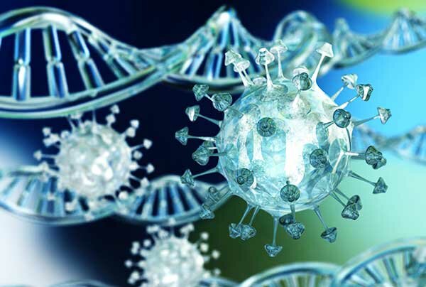
DNA is also known as deoxyribonucleic acid. It is the building block of all life, containing the genetic information that makes each of us an individual. This is known as a genome. A genome is approximately 3,000,000,000 base pairs long and is split into 23 pairs of chromosomes.
DNA gives your body the instructions to create proteins. It is found in the nucleus of all cells and gives the cell the information for normal function, reproduction, and growth. With the exception of identical twins, every human on the planet has a completely unique DNA structure.
This is often used by forensic scientists to help catch criminals. Any DNA left at a crime scene is carefully collected and analyzed. The DNA is extracted from the nuclei and analyzed to create what is known as a DNA profile.
This profile is then compared to a database of other known DNA profiles. This system can pick out correlating DNA sequences and this can be used to identify individuals.

Structure
The structure of DNA is a double helix, formed of 2 strands of a phosphate backbone. This is held together by base pairs to form a kind of ladder shape. This is then twisted to create the classic double helix shape that we all remember from our schooldays.
The phosphate backbone is created from alternating a sugar (the deoxyribose) with phosphate groups. These alternate as phosphate groups cannot attach to one another and need the sugar to act as a glue. The backbone is made intracellularly first, to hold the bases in the correct order.
There are four bases found in DNA. These are known as A, T, G, and C. Their full names are adenine, cytosine, guanine, and thymine. There are specific pairing patterns between these bases.
Adenine and thymine will always bond, and so will cytosine and guanine. These form what is known as a base pair. The order of these pairs dictates the genome’s characteristics. The base pairs are bonded together with hydrogen bonds.
Standard notation of DNA sequences is from 5’ to 3’ (5 prime to 3 prime). This dictates the direction of the elongation of the primer in DNA synthesis. It always goes in the direction of 5’ to 3’.
One side of the backbone will be 5’ to 3’, while the other is the opposite, 3’ to 5’. This is known as an antiparallel running direction. The 5’ to 3’ strand is known as the sense strand, and the other the antisense strand.
Protein synthesis
Each section of 3 base pairs is known as a codon. This dictates which amino acid is created by the body during protein synthesis. There are 20 different amino acids that the body can create, all of which are vital to the creation of proteins.
There are 64 separate codons in the body, but only 61 of these act as coding for amino acids. The other 3 are used to stop more amino acids from being added, and to signal the end of the protein chains.
These instructions for protein synthesis make up only 2% of the human genome. The remaining 98% is known as non-coding DNA.
What is the function of non-coding DNA?
This helps to regulate when the proteins are synthesized in the body. This DNA is also used to control the packaging of the DNA within cells. There are also areas known as scaffold attachment regions, which provide structure to the DNA chain.
Some of this non-coding DNA is transcribed into functional non-coding RNA such as transfer, ribosomal, and regulatory RNA.
Function
The backbone links the nucleotides (base pairs) together in an incredibly stable format. This means that specific enzymes are required to break the bonds as it is very difficult to do.
During DNA synthesis an ATP (adenosine triphosphate) molecule is created. This is an energy molecule that is part of the phosphate backbone and is used to link the DNA.
Replication
This is a process where a cell makes an identical copy of its genome and then divides.
The double helix separates through a process known as unzipping. This pulls apart the 2 phosphate backbone strands. An enzyme known as helicase is used to break the hydrogen bonds between the bases.
This splitting creates a Y shape with the 2 strands of DNA - this is known as a replication fork. Each strand then acts as a kind of template to create a new DNA strand.
The 3’ to 5’ strand (towards the fork of the Y shape) is called the leading strand, and the other is known as the lagging strand. They have different orientations and will replicate in different ways.
A section of RNA produced by the enzyme primase (known as a primer) binds to the end of the leading strand. This serves as the starting point for DNA synthesis.
The enzyme DNA polymerase binds to the leading strand and moves down the length, adding complementary nucleotides bases to the DNA. This strand will be going in the 5’ to 3’ direction. This is known as continuous replication.
The lagging strand will have lots of RNA primers that bind at various points along the length. Okazaki fragments (small chunks of DNA) are added to this lagging strand in a 5’ to 3’ direction. These little fragments will need joining later, therefore this process is known as discontinuous replication.
When all of the bases have been paired up, an enzyme known as exonuclease gets rid of the primers. These empty sections are replaced with further complementary nucleotides.
The strand is then ‘proofread’ to ensure the DNA sequence has been replicated successfully.
Another enzyme, DNA ligase, is used to bind the sequence of DNA into 2 continuous strands.
This will have created 2 molecules of DNA, half new and half old nucleotides. The process of DNA replication is called semi-conservative as only half the DNA molecule is new.
The DNA molecule will then wind up to form a double helix.
